PSMA pathway
PSMA expression in prostate cancer can be mapped with PSMA PET and targeted with lutetium-177–labeled radioligand therapy in appropriately selected patients.
Molecular imaging + targeted therapy
An interactive visual story of how molecular targets and native iodine handling connect diagnostic imaging with radiopharmaceutical treatment—from prostate and neuroendocrine tumors to differentiated thyroid cancer.
The site follows PSMA, somatostatin receptors, and the thyroid cell's sodium–iodide symporter from biologic target to treatment. Imaging, pathology, laboratory data, and clinical context work together to select patients and assess response.
PSMA expression in prostate cancer can be mapped with PSMA PET and targeted with lutetium-177–labeled radioligand therapy in appropriately selected patients.
SSTR expression in well-differentiated neuroendocrine tumors can be visualized with DOTATATE PET and targeted using lutetium-177 DOTATATE peptide receptor radionuclide therapy.
Differentiated thyroid cells can concentrate iodine. In selected patients after thyroidectomy, iodine-131 can ablate remnant tissue or treat iodine-avid residual, recurrent, or metastatic disease.
The sculptural cell image introduces PSMA as a target. A later production version could layer in receptor labels, hover states, or a true 3D turntable without changing the visual hierarchy.

PSMA expression: Many prostate cancers display increased prostate-specific membrane antigen on the cell surface. Expression varies among and within tumors, so this illustration is explanatory—not a quantitative representation.
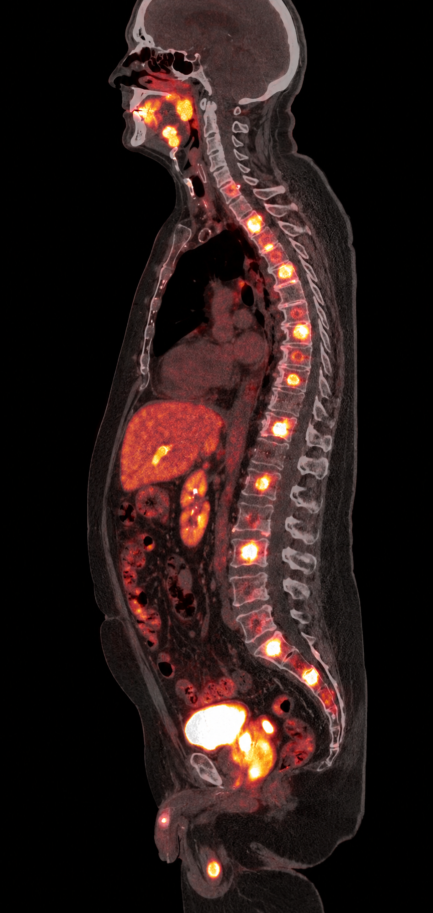
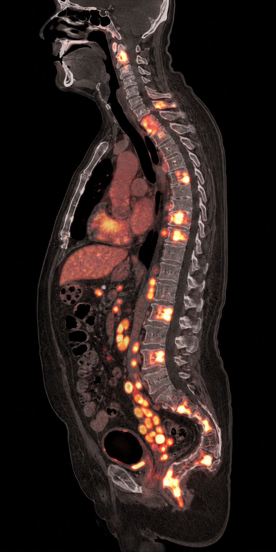
Use the display control to compare PET-emphasized fusion with a grayscale CT-emphasis view, then follow the additional sagittal example to see how PSMA-avid bone disease can appear along the spine.
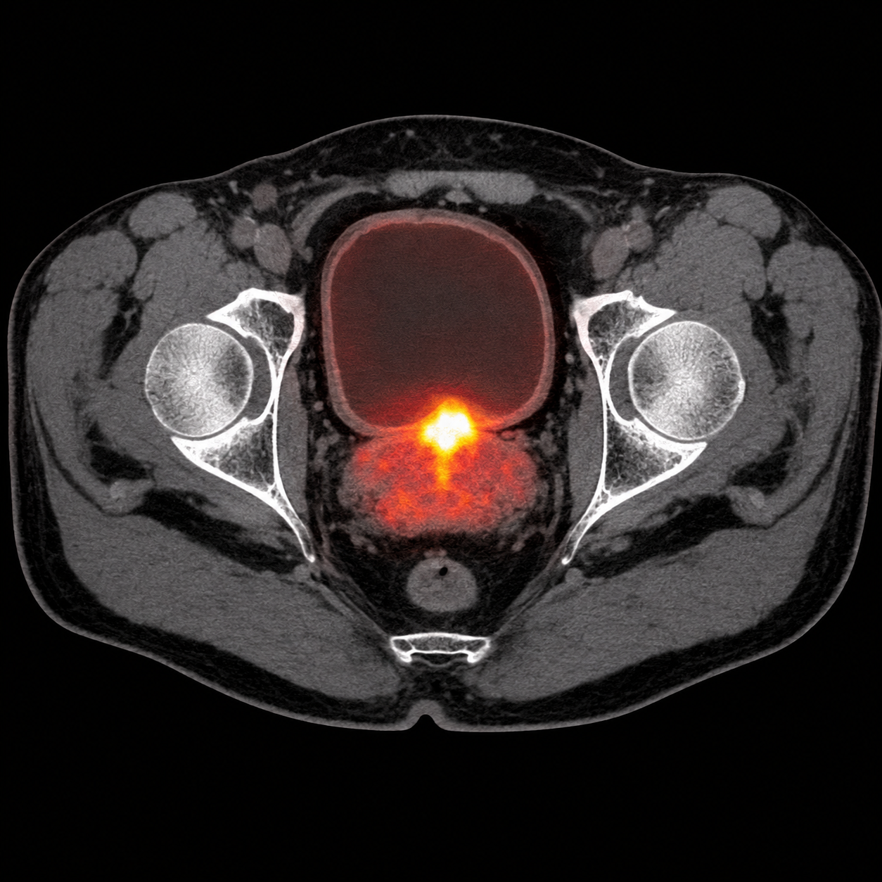
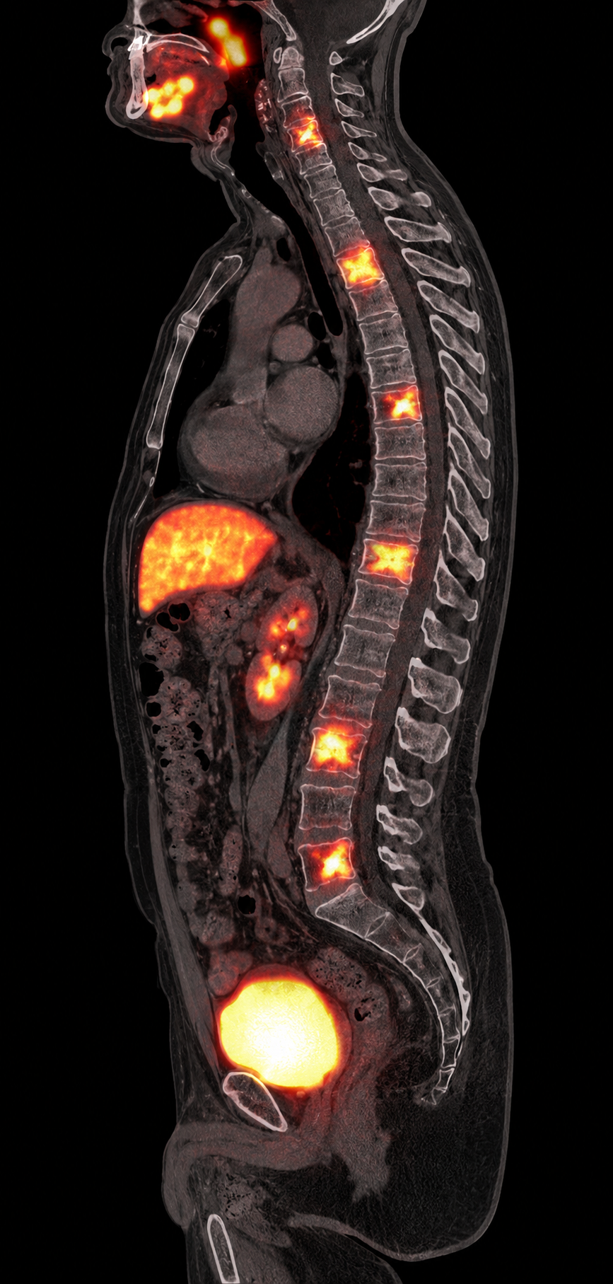
In this teaching example, several bright foci localize to vertebral bodies on the sagittal CT, illustrating multiple PSMA-avid osseous metastases. Expected tracer activity in normal organs—especially the salivary glands, kidneys, liver, and urinary bladder—must be distinguished from disease.
Gallium-68 is a positron emitter used at diagnostic activity for PET. The scanner detects the resulting 511-keV annihilation photons to show where the PSMA-targeted tracer accumulates. Its purpose here is imaging and patient assessment—not delivery of a tumoricidal radiation dose.
Lutetium-177 emits beta particles that deposit energy over a short tissue range and can damage DNA in targeted and nearby tumor cells. It also emits lower-abundance photons that may be used to image treatment distribution, but its principal role is therapy.
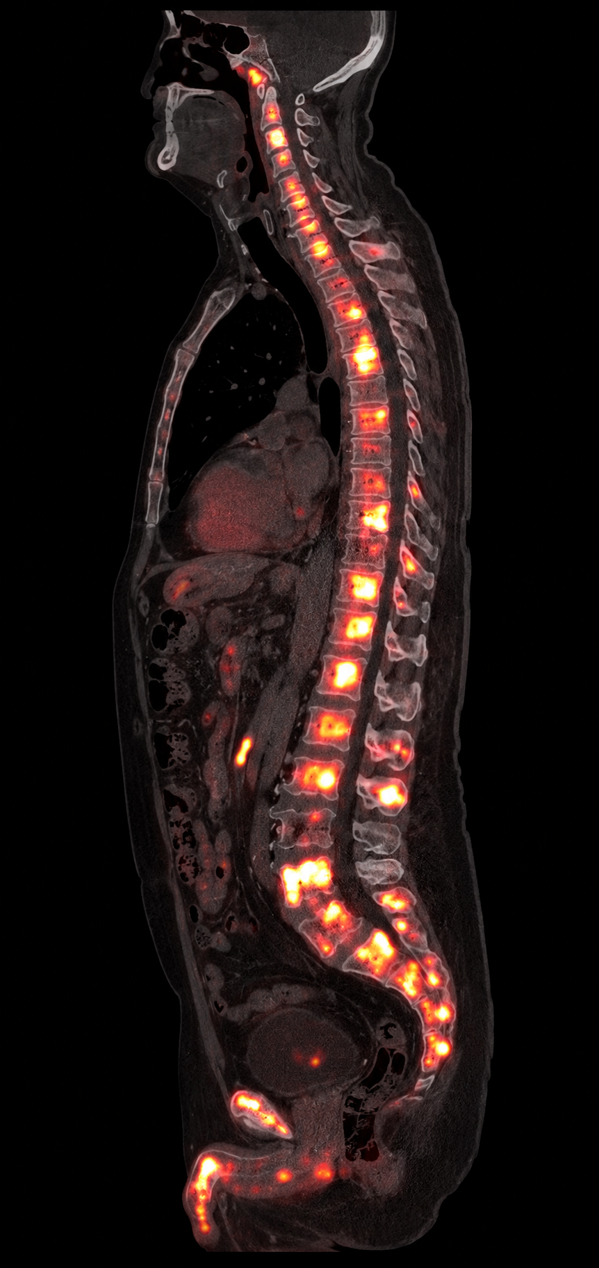
The diagnostic PET/CT identifies PSMA-expressing sites before therapy. After administration, photons emitted by lutetium-177 can be detected with SPECT/CT, allowing the treatment's distribution to be visualized.
The sharper PET signal identifies several PSMA-avid vertebral targets and helps establish whether disease distribution is suitable for PSMA-directed treatment.
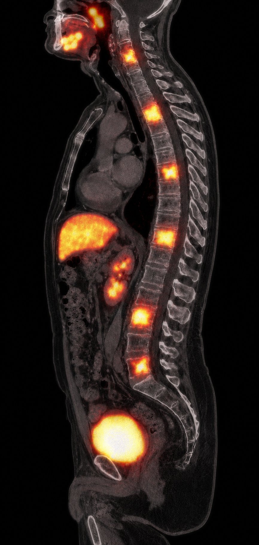
The broader SPECT foci correspond to the pretreatment PET-positive vertebral sites. Beta-minus emissions deliver treatment, while accompanying 113- and 208-keV photons make this post-treatment image possible.
Activity at the same vertebral targets supports successful radioligand delivery and target engagement. It does not, by itself, prove tumor response or clinical efficacy; those require follow-up imaging, laboratory findings, symptoms, and longitudinal assessment.
PSMA PET first helps determine whether disease expresses enough target for PSMA-directed therapy. Then, for selected patients, the radioligand binds, internalizes, and delivers short-range beta radiation.
Lu-177 PSMA therapy depends on the radioligand reaching PSMA-expressing tumor. Greater and more uniform uptake generally signals more available target; absent or markedly heterogeneous uptake can identify disease that may receive less radiation.
Current U.S. prescribing information requires selection with an approved PSMA PET product. Eligibility now spans defined metastatic androgen-pathway-modulation-naïve or -sensitive disease treated with Pluvicto plus an ARPI, as well as mCRPC after ARPI therapy before or after taxane in the labeled settings.
The pivotal VISION trial required at least one PSMA-positive lesion and excluded patients with certain size-qualified PSMA-negative lesions. A strongly avid focus does not compensate automatically for clinically important non-avid disease elsewhere.
Uptake intensity alone does not establish eligibility. Disease setting, prior therapy, laboratory values, organ function, marrow reserve, competing imaging findings, and product-specific criteria also matter.
For oncologists accustomed to chemotherapy, Pluvicto can be viewed as a PSMA-selected systemic anticancer treatment: it circulates intravenously, treats visible and occult PSMA-expressing disease throughout the body, and has its own response pattern, organ monitoring, dose delays, and cumulative marrow considerations.
A fit patient has de novo metastatic prostate cancer, at least one PSMA-positive metastasis on approved PSMA PET, and is beginning systemic hormonal therapy. The aim is to delay radiographic progression before resistant clones and cumulative morbidity dominate.
A patient progresses on abiraterone or enzalutamide, remains taxane-naïve, has PSMA-positive disease, and would benefit from postponing docetaxel because of neuropathy risk, frailty, occupational priorities, or informed preference.
A patient has PSMA-positive progressive mCRPC after an ARPI and one or two taxane regimens, with painful bone or nodal disease, declining tolerance of cytotoxic therapy, but adequate marrow and renal reserve.
“Super responder” is an informal term, not a validated eligibility category. It usually describes an unusually deep and durable biochemical, imaging, and symptomatic response. Favorable signs may include intense, relatively homogeneous PSMA uptake, a large early PSA decline, improving symptoms, preserved counts and renal function, and persistent PSMA expression—but none guarantees duration of benefit.
The FDA-approved initial course remains a maximum of 6 doses. Selected prior responders have received additional cycles or later rechallenge in experienced centers and clinical trials, sometimes using treatment holidays. This can be reasonable to study when disease remains PSMA-avid and marrow, renal, and salivary toxicity are acceptable, but treatment beyond six cycles is not established labeled routine care; prospective trials such as RE-LuPSMA and flexible-schedule studies are testing it.
| Trial | Clinical question | Key finding | Practice message |
|---|---|---|---|
| VISION • phase 3 | Post-ARPI, post-taxane PSMA-positive mCRPC: Pluvicto plus standard care versus standard care. | OS 15.3 vs 11.3 months (HR 0.62); imaging-PFS 8.7 vs 3.4 months (HR 0.40). | A survival-prolonging systemic option—not merely palliation or a salvage scan-directed procedure. |
| PSMAfore • phase 3 | Taxane-naïve mCRPC after one ARPI: Pluvicto versus ARPI switch. | Primary median rPFS 9.3 vs 5.6 months (HR 0.41); substantial crossover limited unadjusted OS interpretation. | Discuss before chemotherapy when the patient is appropriate to delay taxane; avoid automatic ARPI-to-ARPI sequencing. |
| PSMAddition • phase 3 | PSMA-positive metastatic hormone-sensitive disease: Pluvicto plus ADT/ARPI versus ADT/ARPI. | Risk of radiographic progression or death reduced by 28% (HR 0.72); OS trend favored combination but was immature. | Supports early intensification and the July 31, 2026 U.S. combination approval; the studied and approved course is up to six doses. |
| TheraP • randomized phase 2 | Post-docetaxel mCRPC: Lu-PSMA-617 versus cabazitaxel using stringent PSMA/FDG PET selection. | PSA50 response 66% vs 37%; grade 3–4 events 33% vs 53%; mature OS similar. | A credible alternative to cabazitaxel for appropriately imaged patients, with toxicity and quality-of-life differences that matter. |
| ENZA-p • randomized phase 2 | First-line mCRPC: adaptive Lu-PSMA-617 plus enzalutamide versus enzalutamide alone. | Median PSA-PFS 13.0 vs 7.8 months (HR 0.43). | Supports biologic synergy and adaptive dosing concepts; it does not replace the product label for routine sequencing. |
| UpFrontPSMA • randomized phase 2 | De novo high-volume hormone-sensitive disease: two Lu-PSMA-617 cycles followed by docetaxel versus docetaxel alone. | Undetectable PSA at 48 weeks: 41% vs 16%, without an observed increase in severe toxicity. | The “two-cycle” concept belongs to this investigational sequential chemo strategy—not to the FDA-approved Pluvicto-plus-ARPI regimen. |
| LUNAR • randomized phase 2 | Oligorecurrent hormone-sensitive disease with 1–5 PSMA PET-positive lesions: SBRT to all sites versus two neoadjuvant cycles of 177Lu-PNT2002 (6.8 GBq, 6–8 weeks apart) followed by SBRT. | Median PFS was 17.6 versus 7.4 months (HR 0.37); median hormone-therapy-free survival was 24.3 versus 14.1 months. Nearly all progression events were new lesions rather than in-field failures. | Illustrates a systemic-plus-local strategy aimed at occult micrometastatic disease while ablating visible lesions. LUNAR studied PNT2002—not branded Pluvicto—and does not establish two-cycle Pluvicto plus SBRT as routine care. |
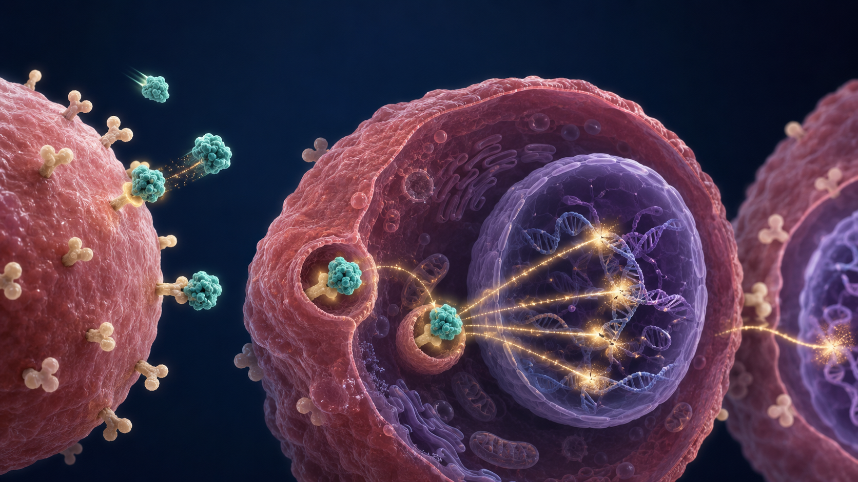
The targeting portion of the radioligand recognizes and binds to PSMA proteins on the cancer-cell surface.
Many well-differentiated neuroendocrine tumors express somatostatin receptors—especially SSTR2. DOTATATE uses that receptor as a molecular address for diagnostic PET and, in selected receptor-positive GEP-NETs, lutetium-177 peptide receptor radionuclide therapy.
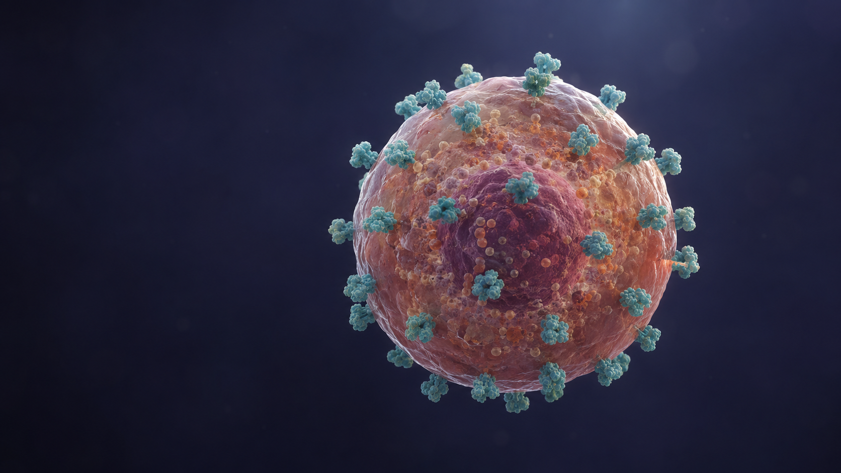
SSTR2 expression: Well-differentiated neuroendocrine tumor cells often retain somatostatin receptors on their surface. Receptor density and distribution vary between patients and lesions, so this illustration explains the target rather than quantifying it.
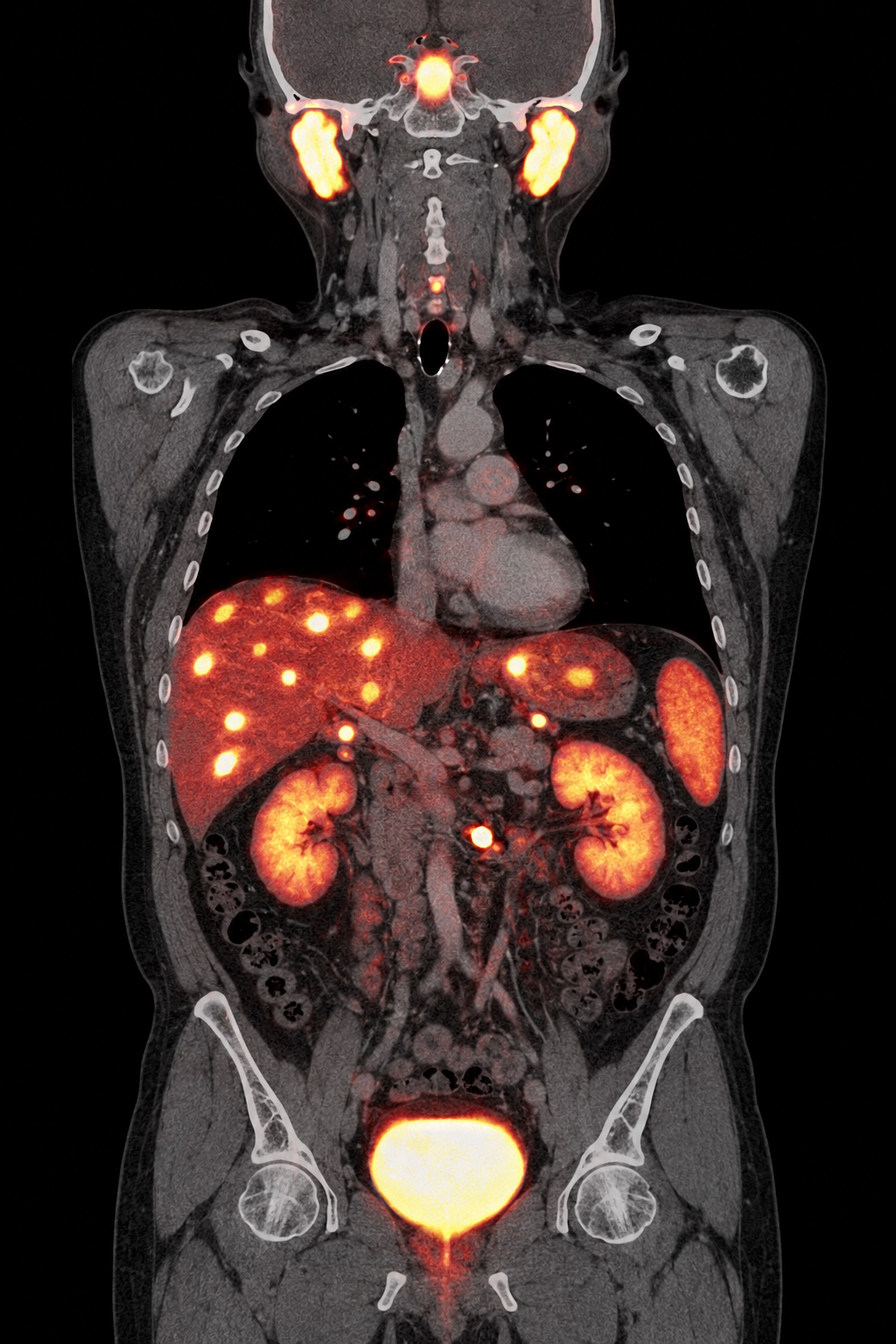
Gallium-68 DOTATATE binds to somatostatin receptors. PET signal therefore reflects receptor distribution—not simply tumor size—and fused CT provides anatomic localization.
Gallium-68 is a positron emitter used for PET localization of somatostatin-receptor-positive NETs. Its diagnostic activity produces a molecular map; it is not administered to deliver a therapeutic tumor dose.
Lutetium-177 DOTATATE binds preferentially to SSTR2, is internalized, and emits beta-minus particles that damage receptor-positive tumor cells and nearby cells. Accompanying photons can also support post-treatment imaging and dosimetry.
PRRT requires sufficient somatostatin-receptor expression for the radiolabeled peptide to localize in tumor. Greater, more uniform uptake generally supports targetability; absent or heterogeneous uptake can indicate disease that may receive less absorbed radiation.
SSTR PET is used to map receptor expression and disease extent. Tumors without somatostatin receptors may not be visualized and are unlikely to be effectively targeted by DOTATATE.
Clinical protocols commonly assess whether tumor uptake is meaningfully greater than normal background organs such as liver. The complete distribution matters more than one exceptionally avid lesion.
Differentiation, grade, disease tempo, prior therapy, renal function, marrow reserve, liver function, symptoms, and receptor heterogeneity all contribute to treatment decisions.
Imaging confirms that the therapeutic target is present, while pathology, disease setting, organ function, prior treatment, and patient goals determine whether lutetium-177 DOTATATE is appropriate.
The Krenning score is a visual, semi-quantitative comparison of tumor uptake with normal reference organs. It was developed for indium-111 pentetreotide scintigraphy; a modified version is commonly applied to SSTR PET. Because PET is more sensitive, scores across modalities are not automatically interchangeable.
Practical interpretation: uptake at least equal to liver (score 2 or higher) has often been used as a minimum proposed threshold, while uptake greater than liver (scores 3–4) gives stronger evidence of SSTR targetability. A single score must not override lesion-to-lesion heterogeneity or the full clinical assessment.
NETTER-1 randomized 229 adults with inoperable, well-differentiated, SSTR-positive midgut NETs progressing on standard-dose octreotide LAR. Patients received four 7.4-GBq cycles of lutetium-177 DOTATATE plus octreotide LAR 30 mg or high-dose octreotide LAR 60 mg.
NETTER-2 randomized 226 patients with newly diagnosed, advanced, well-differentiated grade 2 or grade 3 GEP-NETs (Ki-67 10%–55%) to lutetium-177 DOTATATE plus octreotide LAR 30 mg or octreotide LAR 60 mg.
NETTER-3 is evaluating four cycles of lutetium-177 DOTATATE plus octreotide LAR versus octreotide LAR alone in approximately 240 newly diagnosed patients with SSTR-positive, well-differentiated grade 1 or grade 2 GEP-NETs, Ki-67 below 10%, and high disease burden. The listing included participants age 12 years and older as of April 30, 2026.
Trial criteria are abbreviated for education. The live registry and study team control eligibility, additional protocol-defined criteria may apply, and recruitment status can change.
Functional NETs can continue releasing serotonin or other bioactive hormones during and after PRRT. A somatostatin analog suppresses that secretion while the antitumor response to lutetium-177 DOTATATE develops; it is supportive endocrine therapy, not an antidote to radiation toxicity.
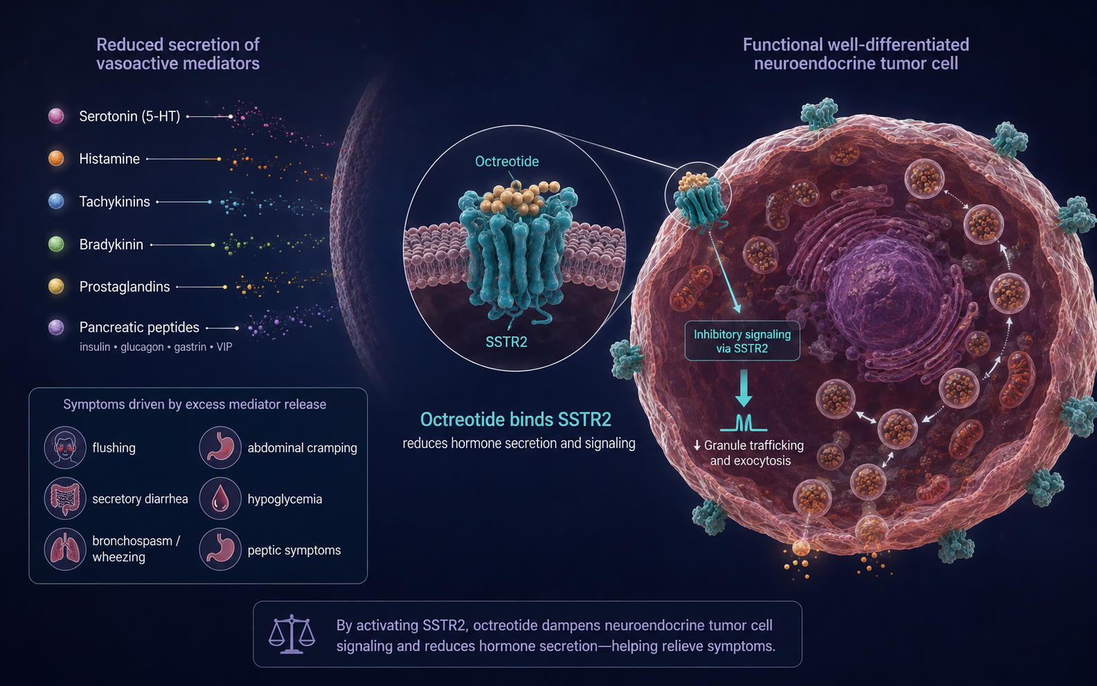
Long-acting octreotide or lanreotide may reduce recurrent flushing, secretory diarrhea, wheezing, hypoglycemia, peptic symptoms, or other syndrome-specific effects, depending on which hormones the tumor produces.
PRRT response is not immediate. Continuing the somatostatin analog helps prevent loss of symptom control between cycles and after the four-cycle course. Short-acting octreotide can be used as rescue therapy when clinically indicated.
Long-acting analogs are held before each Lutathera dose because they may compete for somatostatin receptors. The U.S. label specifies octreotide LAR 30 mg 4–24 hours after each Lutathera dose, then every 4 weeks after the course until progression or 18 months from treatment initiation, at physician discretion.
Octreotide versus lanreotide: the U.S. Lutathera prescribing schedule specifically names octreotide LAR. Lanreotide is also used clinically for NET growth and hormone-syndrome control, but its peri-PRRT timing should follow the treating center's protocol. Short-acting octreotide must be withheld for at least 24 hours, and long-acting somatostatin analogs for at least 4 weeks, before each Lutathera dose unless the treating team directs otherwise.
Side-by-side imaging illustrates the theranostic link: pretreatment DOTATATE PET maps receptor-positive disease, while post-treatment lutetium-177 SPECT shows where the therapeutic radiopharmaceutical localized.
Sharper PET foci show the distribution of SSTR-expressing hepatic metastases and the small abdominal target before therapy.
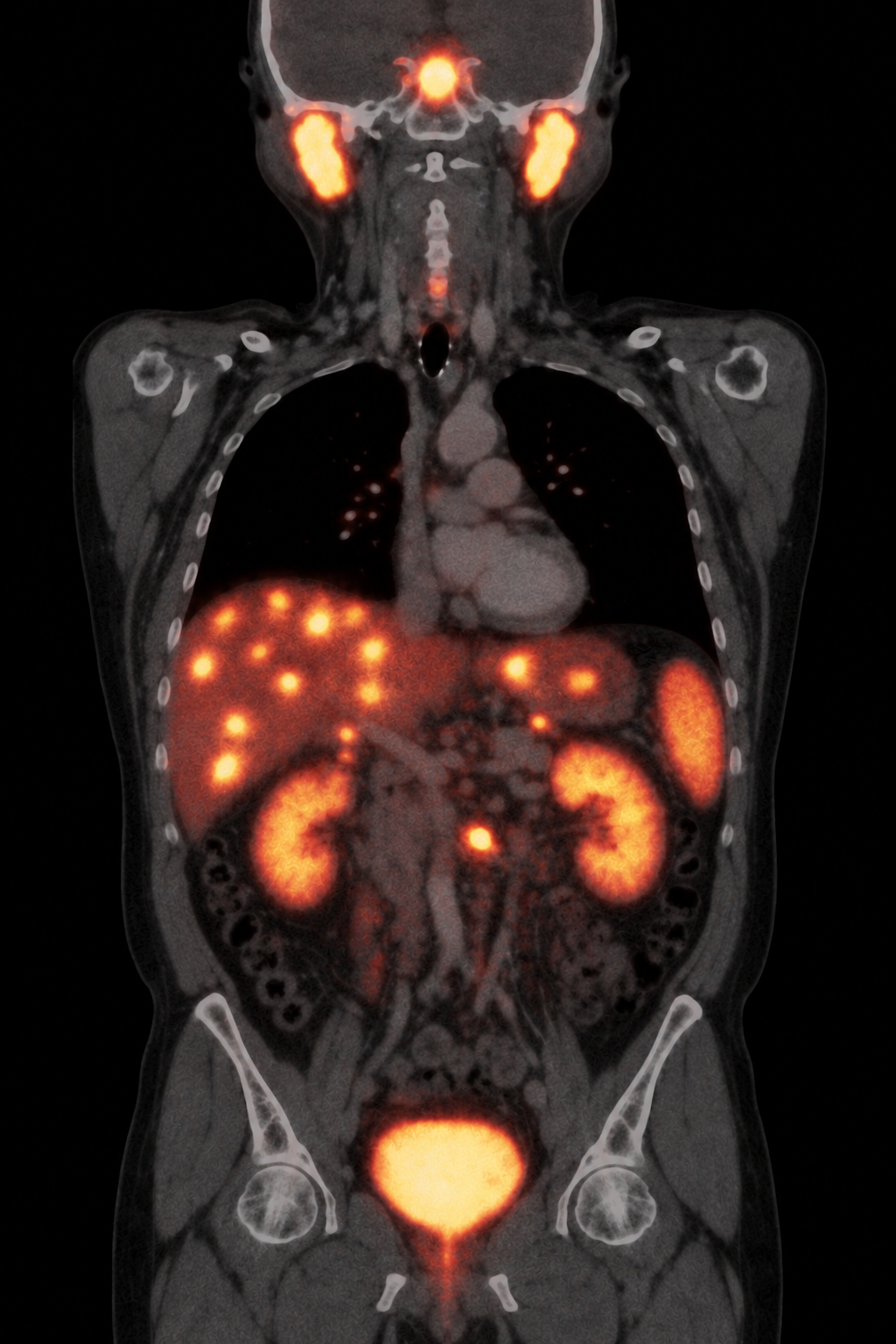
Broader SPECT foci correspond to the PET-positive targets. Concordance supports delivery and target engagement but does not by itself prove tumor response.
DOTATATE provides the receptor-binding component in both examples. Gallium-68 supports diagnostic PET; lutetium-177 supplies therapeutic beta-minus emissions and photons suitable for post-treatment imaging.
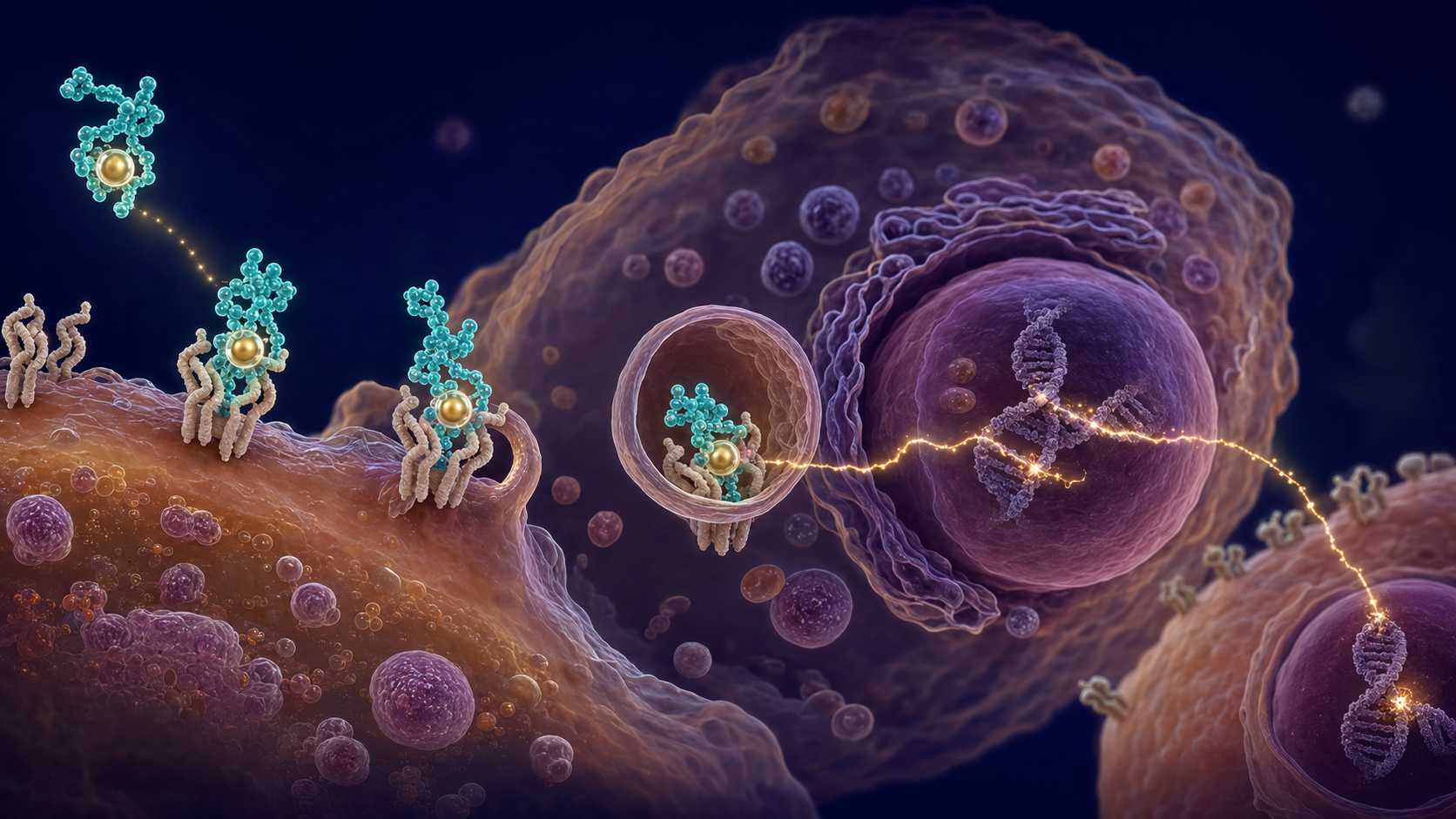
DOTATATE has high affinity for somatostatin receptor subtype 2 on receptor-positive neuroendocrine tumor cells.
Differentiated thyroid cells use the sodium–iodide symporter to concentrate iodide. Iodine-131 follows that native pathway, emitting beta particles for treatment and gamma photons that allow post-therapy whole-body imaging.
Selection begins with histology, surgical findings, postoperative thyroglobulin and anti-thyroglobulin antibodies, neck ultrasound, stage, recurrence risk, prior radioiodine, and evidence that clinically relevant disease retains iodine avidity. Medullary and anaplastic thyroid cancers do not routinely concentrate iodine and are not treated with I-131.
Eliminates residual normal thyroid tissue after total thyroidectomy in selected patients, which may simplify surveillance.
Targets suspected microscopic disease when recurrence risk and expected benefit justify treatment.
Treats iodine-avid residual, recurrent, or metastatic differentiated thyroid cancer when meaningful uptake is expected.
TSH stimulation may use thyroid-hormone withdrawal or recombinant human TSH (Thyrogen), depending on the indication and patient. Medication, supplement, contrast, and dietary iodine exposure are reviewed; the 2025 ATA guideline recommends a low-iodine diet for approximately 1–2 weeks before ablation or treatment.
Review pathology, anatomy, laboratory values, renal function, prior administered activity, pregnancy status, breastfeeding, continence, swallowing, home circumstances, and the patient's ability to follow radiation-safety instructions. Diagnostic I-123 or low-activity I-131 imaging and dosimetry are used selectively.
I-131 is usually given orally. Many patients receive protocol-based activity; lesion or blood/marrow dosimetry may support individualized planning in selected metastatic, pediatric, renally impaired, or previously heavily treated patients. Absorbed dose is not interchangeable with administered activity.
Planar whole-body imaging, often with targeted SPECT/CT, is typically performed several days after treatment. It can reveal additional iodine-avid sites and document distribution, but uptake alone does not establish response.
Time, distance, hygiene, toilet precautions, hydration, and sleeping or travel restrictions are individualized to administered activity, measured exposure, living situation, and regulation. Pregnancy is contraindicated. Breastfeeding must stop well before therapy and must not resume for the current child; written site-specific instructions govern release.
Follow symptoms, examination, TSH-suppressed or stimulated thyroglobulin with antibodies, neck ultrasound, and risk-appropriate anatomic or functional imaging. Rising markers with negative iodine imaging may prompt CT/MRI or FDG PET/CT and evaluation for radioiodine-refractory disease.
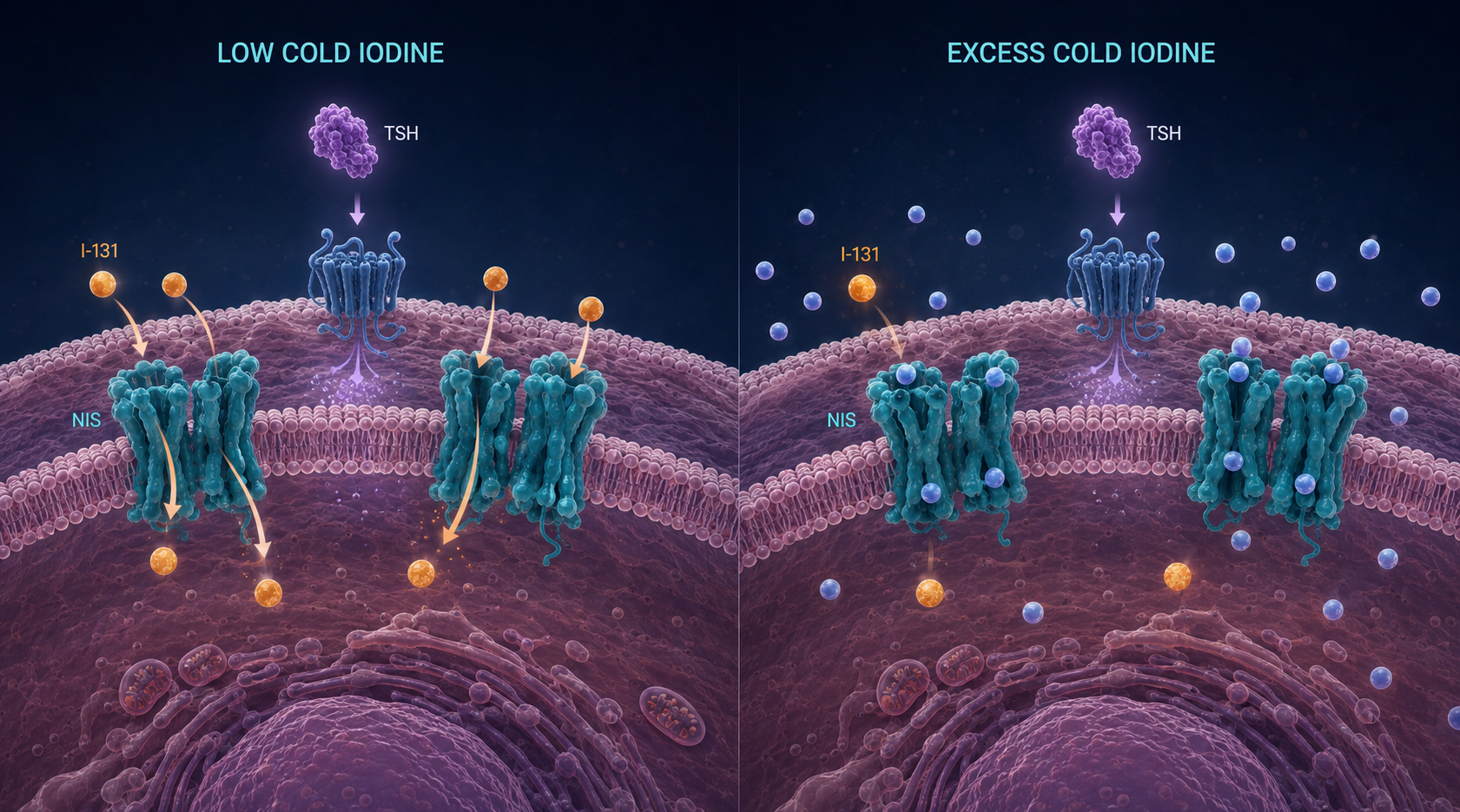
The sodium–iodide symporter (NIS) transports iodide into differentiated thyroid cells. TSH stimulation supports NIS expression and activity, while excess stable “cold” iodine competes with and dilutes I-131, reducing the fraction of radioactive iodine available for uptake.
Two prescribed injections raise TSH while the patient remains on thyroid hormone, usually avoiding prolonged hypothyroid symptoms. It is commonly used for remnant ablation and selected adjuvant treatment; its role in distant metastatic disease requires individualized judgment.
Levothyroxine is stopped according to the treatment protocol so endogenous TSH rises, often targeting a TSH above 30 mIU/L. This may cause fatigue, cognitive slowing, constipation, cold intolerance, or mood symptoms while hypothyroid.
For about 1–2 weeks, avoid major iodine sources such as iodized salt, seaweed, many seafoods, and iodine-containing supplements as directed. Review recent iodinated contrast and amiodarone; do not restrict non-iodized salt unless instructed.
A post-therapy planar whole-body scan is generally obtained 2–10 days after I-131. Focal uptake in the anterior midline thyroid bed commonly represents residual normal thyroid tissue (a thyroid remnant); depending on pathology and location, uptake can also represent residual iodine-avid disease. I-131 is intended to ablate the remnant and treat iodine-avid cancer.
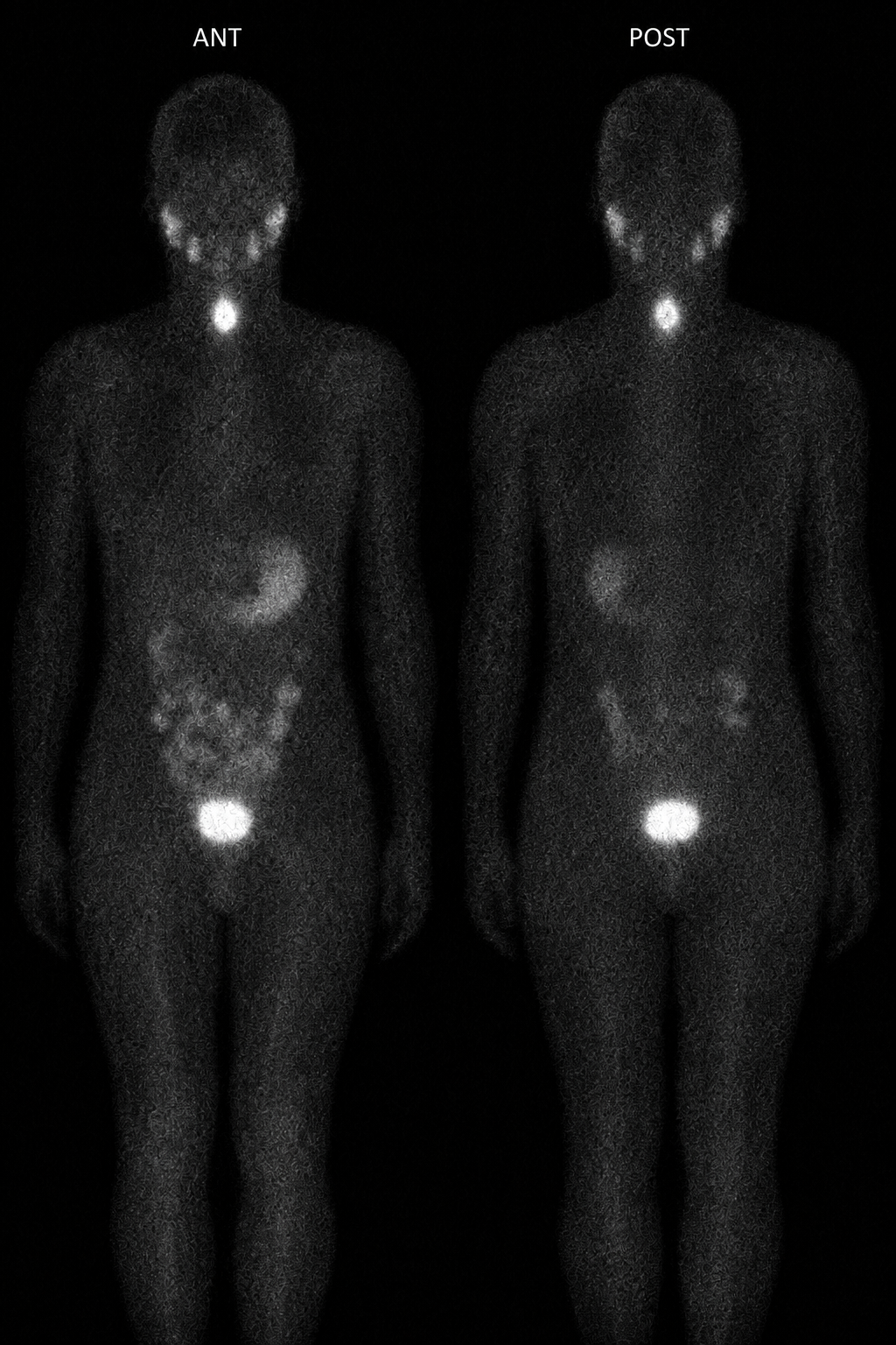
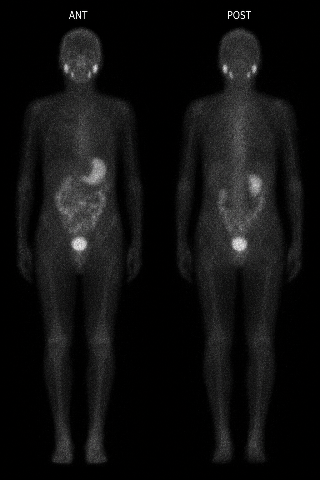
Simulated teaching images—not patient studies. Physiologic salivary, gastric, bowel, and urinary activity can occur. A negative follow-up scan does not by itself prove that no disease remains.
The newest ATA system has four recurrence-risk tiers: low, low-intermediate, intermediate-high, and high. The table preserves the familiar low/intermediate/high summary while showing the updated intermediate subdivisions.
| ATA recurrence risk | Typical RAI approach | Suggested I-131 activity | Therapeutic goal |
|---|---|---|---|
| Low (<10% recurrence) | Usually no RAI. If remnant ablation is chosen after shared decision-making, use the lowest effective activity. | 30–50 mCi (1.1–1.85 GBq) | No treatment or remnant ablation |
| Low-intermediate (10–15%) and intermediate-high (16–30%) | Consider RAI; expected benefit varies with adverse histology, nodal burden/location, postoperative thyroglobulin, age, imaging, and patient priorities. | 30–100 mCi (1.1–3.7 GBq) | Remnant ablation ± adjuvant treatment |
| High (>30%) | RAI is typically recommended after total thyroidectomy when disease is expected to be iodine-avid. | 100–150 mCi (3.7–5.55 GBq) | Remnant ablation and adjuvant treatment |
| Known distant metastases | Treat iodine-avid disease; select empirical activity or individualized dosimetry, especially in advanced age, diffuse pulmonary metastases, renal impairment, or when higher activities are contemplated. | 100–200 mCi (3.7–7.4 GBq), or dosimetry | Treatment of known disease plus remnant ablation |
These are guideline ranges, not prescriptions. Final activity depends on treatment intent, pathology, residual disease, iodine avidity, renal and marrow considerations, prior I-131 exposure, dosimetry when appropriate, patient preference, and multidisciplinary review. The 2025 ATA guideline recommends dosimetry when activities above 200 mCi are contemplated.
Compare how selection, preparation, treatment, dosimetry, safety, and response assessment differ across Lu-177 PSMA, Lu-177 DOTATATE, and I-131. Choose a pathway below.
Each pathway requires an authorized treatment team, product- and patient-specific evaluation, clear radiation-safety counseling, and coordinated oncology follow-up.
Confirm the product-specific prostate-cancer indication and PSMA-positive disease on an approved PET agent; assess non-avid disease, prior therapy, performance status, marrow reserve, and renal function.
Review CBC and renal/hepatic tests, medications, hydration, continence, pain needs, and radiation-safety logistics. Coordinate systemic therapy according to the oncology plan.
Administer Lu-177 PSMA intravenously in cycles. Post-treatment imaging can confirm distribution; quantitative SPECT/CT dosimetry is increasingly used but is not identical across centers.
Use written distance, hygiene, hydration, toilet, travel, and contact precautions. Monitor for myelosuppression, renal toxicity, dry mouth, nausea, and fatigue.
Trend symptoms, performance status, PSA, CBC, renal function, and interval CT/MRI, bone, or PSMA PET imaging when clinically indicated; assess cumulative toxicity before each cycle.
Confirm SSTR-positive, well-differentiated NET in the appropriate disease setting; integrate grade, growth rate, symptoms, FDG/SSTR heterogeneity, marrow reserve, and renal/hepatic function.
Coordinate somatostatin-analog timing, antiemetics, laboratory testing, venous access, and renal-protective amino-acid infusion. Review pregnancy and radiation-safety status.
Administer Lu-177 DOTATATE intravenously in planned cycles with concurrent amino acids. Post-treatment SPECT/CT confirms localization and can support kidney, marrow, and tumor dosimetry.
Apply written radiation precautions and monitor nausea, electrolyte effects, cytopenias, renal/hepatic toxicity, and rare delayed marrow neoplasms. In functional tumors, resume coordinated somatostatin-analog therapy to maintain hormonal symptom control; it is not a treatment for radiation toxicity.
Assess symptoms and hormone-related syndromes, including rescue octreotide needs, plus CBC and organ function; then use CT/MRI and selected SSTR or FDG PET. Morphologic response may lag behind clinical benefit.
Define ablation, adjuvant, or treatment intent using histology, surgery, risk, thyroglobulin/antibodies, ultrasound and cross-sectional imaging, prior I-131, and expected iodine avidity.
Stimulate TSH, reduce competing stable iodine, check pregnancy and breastfeeding status, review renal function and home logistics, and choose empiric or dosimetry-guided activity.
Administer oral I-131. Obtain post-therapy whole-body imaging, with SPECT/CT when useful, to map treatment distribution and refine staging.
Give individualized time-distance, household, hygiene, toilet, hydration, travel, and reproductive instructions. Monitor salivary, lacrimal, marrow, pulmonary, and gonadal risks as relevant.
Use dynamic risk stratification with thyroglobulin/antibodies, TSH management, neck ultrasound, and risk-directed imaging. Identify persistent disease and loss of iodine avidity.
Dosimetry note: Administered activity (MBq or mCi) is the amount given; absorbed dose (Gy) is energy deposited in tissue. Imaging-based dosimetry estimates absorbed dose but methods, timing, calibration, and clinical thresholds vary by therapy and institution.